Diffusion Through a Membrane Lab: Answer Key Insights
The diffusion through a membrane lab explores selectively permeable membranes. It is demonstrated with dialysis tubing acting as a semi-permeable barrier. This reveals diffusion principles, osmosis, and passive transport. Observing red onion cells with salt solution is also key.
The process of diffusion is a fundamental concept in biology, especially when considering how substances move within and between cells. This movement is often facilitated or restricted by membranes, which act as selective barriers. The “Diffusion Through a Membrane” lab is designed to explore these principles in a hands-on manner, providing students with a tangible understanding of abstract concepts.
At its core, diffusion is the net movement of molecules from an area of high concentration to an area of low concentration. This movement is driven by the inherent kinetic energy of molecules, which causes them to randomly move and collide. When there is a concentration gradient, this random movement results in a net flow down the gradient until equilibrium is reached.
Membranes, particularly those found in biological systems, play a crucial role in regulating diffusion. They are not simply passive barriers; instead, they are selectively permeable, meaning that they allow some substances to pass through while blocking others. This selectivity is based on factors such as size, charge, and solubility of the molecules, as well as the properties of the membrane itself. The lab helps illuminate these principles.
Key Concepts: Diffusion, Osmosis, and Passive Transport
Understanding diffusion, osmosis, and passive transport is essential for comprehending how cells maintain their internal environment and interact with their surroundings. These processes are all forms of passive transport, meaning they do not require the cell to expend energy. Instead, they rely on the inherent kinetic energy of molecules and the concentration gradients across membranes.
Diffusion, as previously mentioned, is the movement of molecules from an area of high concentration to an area of low concentration. This applies to various substances, including gases, liquids, and solutes. The rate of diffusion is influenced by factors such as temperature, concentration gradient, and the size and properties of the molecules involved.

Osmosis is a specific type of diffusion that focuses on the movement of water across a semi-permeable membrane. Water moves from an area of high water concentration (low solute concentration) to an area of low water concentration (high solute concentration). This movement is driven by the difference in water potential across the membrane. The experiment showcases how these principles work.
Passive transport encompasses both diffusion and osmosis, along with other processes such as facilitated diffusion. Facilitated diffusion involves the use of transport proteins to assist the movement of molecules across the membrane. Though it requires a protein, it is still passive.
Understanding Semi-Permeable Membranes
Semi-permeable membranes, also known as selectively permeable membranes, are critical components in biological systems. They play a vital role in regulating the movement of substances into and out of cells, organelles, and other compartments. These membranes possess a unique structure that allows some molecules to pass through while restricting the passage of others, based on factors like size, charge, and solubility.
The key characteristic of a semi-permeable membrane lies in its ability to discriminate between different molecules. Typically, small, nonpolar molecules can readily diffuse across the membrane, whereas larger, polar molecules and ions face more difficulty or require assistance from transport proteins. This selective permeability is crucial for maintaining the proper internal environment of cells and for carrying out various cellular processes.
The structure of the cell membrane, composed of a phospholipid bilayer with embedded proteins, is responsible for its semi-permeable nature. The hydrophobic core of the lipid bilayer prevents the passage of charged or polar molecules, while the proteins can act as channels or carriers to facilitate the transport of specific substances. Dialysis tubing serves as a simplified model, exhibiting comparable selective properties.
The understanding of semi-permeable membranes is fundamental to comprehending cellular transport mechanisms and their significance in maintaining cellular homeostasis.
The Role of Solute and Solvent in Diffusion
In the process of diffusion, both the solute and solvent play distinct but interconnected roles. The solute refers to the substance that is dissolved in a liquid, while the solvent is the liquid that dissolves the solute. Understanding their interplay is essential for comprehending how diffusion occurs, especially across membranes. The concentration gradient of the solute drives the diffusion process, with solute molecules moving from an area of high concentration to an area of low concentration until equilibrium is reached.
The solvent, often water in biological systems, provides the medium in which the solute molecules can move. The properties of the solvent, such as its polarity and viscosity, can influence the rate of diffusion. Smaller solvent molecules generally facilitate faster diffusion rates. Furthermore, the interactions between solute and solvent molecules can affect the diffusion process. Solvation, the process by which solvent molecules surround solute molecules, can alter the effective size and mobility of the solute.
Across a semi-permeable membrane, the movement of both solute and solvent is critical. If the membrane is permeable to both, both will diffuse down their concentration gradients until equilibrium. However, if the membrane is only permeable to the solvent, osmosis occurs, where the solvent moves to equalize solute concentrations. Thus, the interplay of solute and solvent dictates the direction and extent of diffusion.
Hypertonic, Hypotonic, and Isotonic Solutions Explained
Understanding hypertonic, hypotonic, and isotonic solutions is crucial when studying diffusion and osmosis, especially in the context of cellular environments. These terms describe the relative solute concentrations of two solutions separated by a semi-permeable membrane. A hypertonic solution has a higher solute concentration compared to another solution, causing water to move out of the other solution and into the hypertonic one via osmosis.
Conversely, a hypotonic solution has a lower solute concentration compared to another solution. In this scenario, water will move from the hypotonic solution into the other solution, attempting to equalize the solute concentrations. This influx of water can cause cells to swell and potentially burst if they lack a rigid cell wall to withstand the pressure.
An isotonic solution has an equal solute concentration compared to another solution. In this case, there is no net movement of water between the two solutions, as the osmotic pressure is balanced. Cells placed in an isotonic solution will neither gain nor lose water, maintaining their normal shape and function. These concepts are fundamental in understanding how cells maintain homeostasis and respond to changes in their surrounding environment.
Membrane Permeability: Factors and Influences
Membrane permeability, the extent to which a membrane allows substances to pass through, is a critical factor in biological processes. Several factors influence membrane permeability, including the size, charge, and polarity of the molecules attempting to cross the membrane. Small, nonpolar molecules generally diffuse across membranes more easily than large, polar ones.
The composition of the membrane itself also plays a significant role. Membranes are primarily composed of a phospholipid bilayer, and the properties of the phospholipids, such as the length and saturation of their fatty acid tails, can affect membrane fluidity and permeability. Cholesterol, another component of cell membranes, also influences fluidity and can reduce permeability to small molecules.
Temperature affects membrane permeability, with higher temperatures generally increasing fluidity and permeability. The presence of transport proteins within the membrane can also significantly alter permeability by facilitating the movement of specific molecules across the membrane. Furthermore, the concentration gradient of a substance across the membrane influences its rate of diffusion, with steeper gradients leading to faster diffusion rates. Understanding these factors is essential for comprehending how cells regulate the movement of substances in and out, maintaining cellular homeostasis.
The Diffusion Through a Membrane Lab: Overview
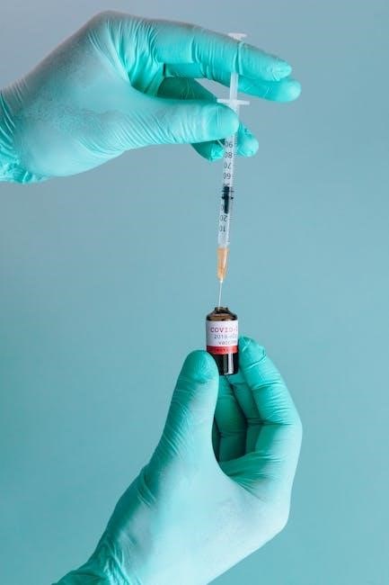
The Diffusion Through a Membrane Lab is a fundamental experiment designed to illustrate the principles of diffusion and osmosis across a semi-permeable membrane. Typically, the lab involves using dialysis tubing, which acts as a model cell membrane, to separate solutions with varying solute concentrations.

The core objective is to observe and analyze the movement of molecules, such as glucose, starch, and iodine (often in the form of a starch indicator solution), across the membrane. Students carefully set up controlled experiments, placing different solutions inside and outside the dialysis tubing and monitoring changes in color or concentration over time. These changes provide evidence of molecular movement driven by concentration gradients.
Data collection involves recording initial and final conditions, noting any color changes, and potentially measuring the mass or volume changes of the solutions. By analyzing this data, students can determine which molecules were able to pass through the membrane and draw conclusions about the membrane’s permeability. The lab reinforces concepts like selective permeability, diffusion, osmosis, and the influence of solute size on molecular movement. It allows students to visualize abstract concepts and develop critical thinking skills in experimental design and data interpretation, all crucial for understanding cellular transport mechanisms.
Dialysis Tubing as a Model Membrane
Dialysis tubing serves as an excellent model for a biological membrane in diffusion experiments due to its semi-permeable nature. This means it allows some molecules to pass through while restricting others, mimicking the selective permeability of cell membranes. The tubing is typically made of cellulose, which has microscopic pores of a specific size.
These pores dictate which molecules can diffuse across the membrane. Smaller molecules, like water, glucose, and ions, can easily pass through the pores, while larger molecules, such as starch and proteins, are too large to permeate. This size-selective permeability is a key characteristic that makes dialysis tubing a useful model.
In a diffusion experiment, the dialysis tubing is usually filled with a solution containing various solutes and then immersed in another solution with different solute concentrations. The movement of molecules across the tubing can then be observed and measured. For example, if the tubing contains glucose and is placed in water, the glucose will diffuse out of the tubing into the water until equilibrium is reached. The rate and extent of this diffusion depend on factors such as the concentration gradient, temperature, and the size of the molecules relative to the pore size of the dialysis tubing. This allows for controlled investigation of key diffusion principles.
Selectively Permeable Membranes in Action
Selectively permeable membranes, like those found in living cells, are crucial for maintaining cellular homeostasis. These membranes allow certain molecules to pass through while restricting others, a process vital for nutrient uptake, waste removal, and maintaining proper internal conditions. This selective permeability is primarily determined by the membrane’s structure, which includes a phospholipid bilayer with embedded proteins.
The phospholipid bilayer itself is generally permeable to small, nonpolar molecules, such as oxygen and carbon dioxide, which can readily diffuse across the membrane. However, it is impermeable to larger, polar molecules and ions, which require the assistance of membrane proteins to cross. These proteins act as channels or carriers, facilitating the transport of specific molecules across the membrane.
In the diffusion through a membrane lab, the selective permeability of the membrane can be observed by using different solutes and solvents. For instance, a membrane may allow glucose to pass through but not starch, demonstrating its ability to discriminate based on size. Similarly, the presence of specific transport proteins can enable the selective uptake of certain ions or nutrients. Understanding how selectively permeable membranes function is essential for comprehending many biological processes, including nerve impulse transmission, muscle contraction, and kidney function. This selective action ensures cellular survival.
Lab Report Essentials: Data Collection and Analysis
A comprehensive lab report is crucial for documenting and interpreting the findings of a diffusion through a membrane experiment. Accurate data collection is the foundation of any strong scientific report. Begin by meticulously recording all initial conditions, such as the concentrations of solutions, the type of membrane used (e.g., dialysis tubing), and the volumes of solutions involved; During the experiment, carefully observe and record any changes occurring within the system, such as color changes, volume alterations, or the presence of specific substances.
Quantitative data, such as mass or volume measurements taken at regular intervals, should be organized into tables for easy analysis. Qualitative observations, like color changes, should be described in detail, noting the timing and intensity of the changes. Data analysis involves calculating rates of diffusion, comparing experimental results to controls, and identifying any trends or patterns in the data. Graphs can be used to visualize the data and make it easier to identify trends.
Statistical analysis may be necessary to determine the significance of the results and to rule out the possibility that the observed effects are due to chance. Be sure to properly label all axes on graphs and include legends for clarity. The final step involves interpreting the data in the context of the experimental hypothesis and discussing any potential sources of error or limitations of the study. The lab report will reveal the experiment details.
Observing Changes in Red Onion Cells with Salt Solution
Observing red onion cells when exposed to a salt solution provides a clear visual demonstration of osmosis and its effects on cellular structures. Red onion cells are ideal for this experiment because their pigmented vacuoles make it easy to observe changes within the cell. When a red onion cell is placed in a hypertonic salt solution, which has a higher solute concentration than the cell’s interior, water moves out of the cell and into the surrounding solution via osmosis.
This water movement causes the cell membrane to shrink away from the cell wall, a phenomenon known as plasmolysis. The cell’s contents appear to contract, and the normally turgid cell becomes flaccid. The degree of plasmolysis is dependent on the concentration of the salt solution; higher concentrations result in more pronounced effects.
To observe these changes, prepare a wet mount of red onion cells in distilled water as a control. Then, prepare another wet mount with a drop of salt solution. Compare the appearance of the cells under a microscope. Note the cell membrane pulling away from the cell wall in the salt solution, indicating water loss. This experiment visually reinforces the concept of osmosis and how solute concentrations affect water movement across cell membranes. The changes are easily visible.
Osmosis: Diffusion of Water Across a Membrane
Osmosis is a specific type of diffusion that focuses solely on the movement of water molecules across a semi-permeable membrane. This membrane allows water to pass through but restricts the passage of certain solutes. The driving force behind osmosis is the difference in water potential between two solutions separated by the membrane. Water potential is influenced by solute concentration and pressure; water moves from an area of high water potential (low solute concentration) to an area of low water potential (high solute concentration).
In biological systems, osmosis is crucial for maintaining cell turgor, regulating fluid balance, and facilitating nutrient uptake. For example, plant cells rely on osmosis to maintain their rigidity, which is essential for structural support. Animal cells, on the other hand, must carefully regulate osmosis to prevent swelling or shrinking due to water influx or efflux.
Understanding osmosis requires considering the concepts of hypertonic, hypotonic, and isotonic solutions. Water moves from hypotonic solutions (lower solute concentration) to hypertonic solutions (higher solute concentration) until equilibrium is reached, resulting in isotonic conditions where the solute concentrations are equal on both sides of the membrane. The principles of osmosis underpin many physiological processes and are vital for life.

Starch Indicator Solution and Membrane Permeability
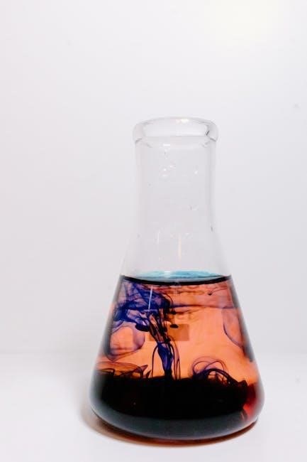
In the context of a diffusion through a membrane lab, a starch indicator solution plays a crucial role in determining the permeability of the membrane to starch molecules. Typically, iodine or Lugol’s solution is used as the starch indicator. This solution reacts with starch to produce a distinct color change, usually turning from a pale yellow or brown to a deep blue-black color. This color change indicates the presence of starch.
The experiment involves placing the starch indicator solution on one side of a semi-permeable membrane, such as dialysis tubing, and a starch solution on the other side. By observing whether the starch indicator solution changes color over time, one can deduce whether starch molecules have crossed the membrane. If the indicator solution turns blue-black, it indicates that starch has diffused through the membrane and reacted with the indicator.
If no color change occurs, it suggests that the membrane is impermeable to starch molecules, meaning that the pores in the membrane are too small for starch to pass through. This experiment effectively demonstrates the concept of selective permeability, where membranes allow certain molecules to pass while restricting others based on size and other properties. The results provide valuable insights into the membrane’s characteristics.
Experiment Setup: Demonstrating Molecular Movement
Setting up an experiment to demonstrate molecular movement across a membrane involves careful consideration of materials and procedures. Typically, dialysis tubing is used as a model semi-permeable membrane. This tubing has microscopic pores that allow small molecules to pass through while restricting larger ones. The experiment often includes solutions of varying solute concentrations, such as glucose, starch, or salt, placed on either side of the membrane.
To begin, the dialysis tubing is prepared by soaking it in water to soften it. One end is then clamped or tied off securely. A solution containing one or more solutes is poured into the open end of the tubing, and the other end is similarly sealed, creating a small bag. This bag is then submerged in a beaker containing a different solution. The beaker solution may contain different solutes or simply be distilled water.
Over time, molecules will move across the membrane due to diffusion and osmosis. The movement of these molecules can be tracked by periodically sampling the solutions on either side of the membrane and testing for the presence of specific solutes. For instance, a glucose test strip can detect glucose, while a starch indicator solution can detect starch. By monitoring these changes, students can observe and quantify the movement of molecules across the membrane, providing a clear demonstration of diffusion principles.
Common Errors and Troubleshooting in the Lab

Several common errors can occur during a diffusion through a membrane lab, affecting the accuracy of results. One frequent mistake is improper preparation of the dialysis tubing. If the tubing is not thoroughly soaked, it may not be sufficiently permeable, hindering molecular movement. Another error involves incorrect sealing of the tubing, leading to leakage and cross-contamination of solutions. Ensuring a tight seal is crucial for maintaining the integrity of the experiment.
Another potential issue is using incorrect concentrations of solute solutions. If the concentrations are too similar, the rate of diffusion may be too slow to observe significant changes within the allotted time. Conversely, excessively high concentrations can lead to osmotic pressure issues and potential bursting of the dialysis tubing.
Contamination of samples is another concern. Using dirty glassware or accidentally introducing foreign substances can skew results. Always use clean equipment and follow proper handling procedures. When testing for the presence of specific solutes, such as glucose or starch, ensure that the testing reagents are fresh and properly stored. Incorrect reagent preparation or expired reagents can lead to false positives or negatives.
If unexpected results occur, carefully review the experimental setup and procedures to identify potential sources of error. Repeating the experiment with meticulous attention to detail can often resolve discrepancies and provide more accurate data.
Analyzing Results: Glucose, Starch, and Starch Indicator
Analyzing the results of a diffusion through a membrane lab often involves assessing the presence or absence of glucose, starch, and starch indicator solution (iodine) in different compartments. The key lies in understanding their molecular sizes and the membrane’s permeability.

Typically, glucose, being a small molecule, should be able to diffuse across the membrane from a high concentration area to a low concentration area. If glucose is initially inside the dialysis tubing, testing the surrounding water for glucose will indicate whether diffusion occurred. A positive test confirms glucose passage.
Starch, a large polysaccharide, is generally too large to pass through the pores of the dialysis tubing. Therefore, if starch is initially inside the tubing, it should remain there throughout the experiment. Testing the surrounding water should yield negative results for starch.
The starch indicator, iodine solution, is used to detect the presence of starch. When iodine comes into contact with starch, it forms a dark blue or black complex. If iodine is initially outside the dialysis tubing and starch is inside, the iodine may diffuse into the tubing. If starch is present inside, the solution within the tubing will turn dark blue or black, indicating that the iodine has diffused in and reacted with the starch.
Conversely, if iodine is inside and starch is outside, a blue-black color change in the external solution would suggest membrane compromise.
Reinforcing Key Diffusion Principles
Interpreting Data: Identifying Unknown Solutions
Interpreting data from a diffusion through a membrane lab to identify unknown solutions relies on understanding the principles of diffusion, osmosis, and selective permeability. Dialysis tubing, acting as a model membrane, allows for the separation of molecules based on size.
Begin by carefully observing any color changes, or lack thereof, within the dialysis bag and the surrounding solution. If iodine is present, the appearance of a dark blue or black color indicates the presence of starch, as the iodine reacts with starch to form a colored complex. If the solution remains clear, starch is likely absent.
Next, test the solutions inside and outside the dialysis bag for the presence of glucose using a glucose test strip. A positive result suggests that glucose molecules have diffused across the membrane. Comparing the concentrations of glucose inside and outside the bag can provide insight into the initial concentration gradients.
Consider the movement of water across the membrane. If the dialysis bag has gained weight, water has entered due to osmosis, indicating that the solution inside the bag was hypertonic relative to the surrounding solution. Conversely, a loss of weight suggests water has exited the bag, indicating a hypotonic internal environment.

By analyzing the diffusion patterns of known substances and observing osmotic changes, one can deduce the identity of unknown solutions based on their molecular composition and concentration.